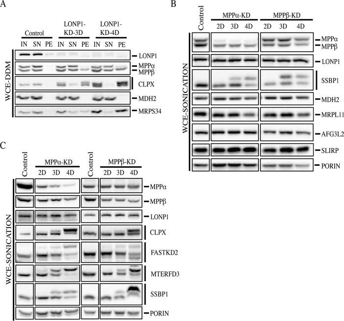
FIG 7.
Analysis of MPP-depleted cell lines. (A) Input, supernatant, and pellet fractions from whole-cell extracts treated with n-dodecyl-β-d-maltoside were used for SDS-PAGE immunodetection of the indicated proteins in control and LONP1-depleted cells (3 and 4 days). (B and C) Immunoblot analysis of whole-cell extracts obtained by sonication from control cells, MPPα-depleted cells (MPPα-KD), and MPPβ-depleted cells (MPPβ-KD) for 2, 3, and 4 days. Immunoblots show the steady-state levels of the indicated proteins. Porin was used as a loading control.

