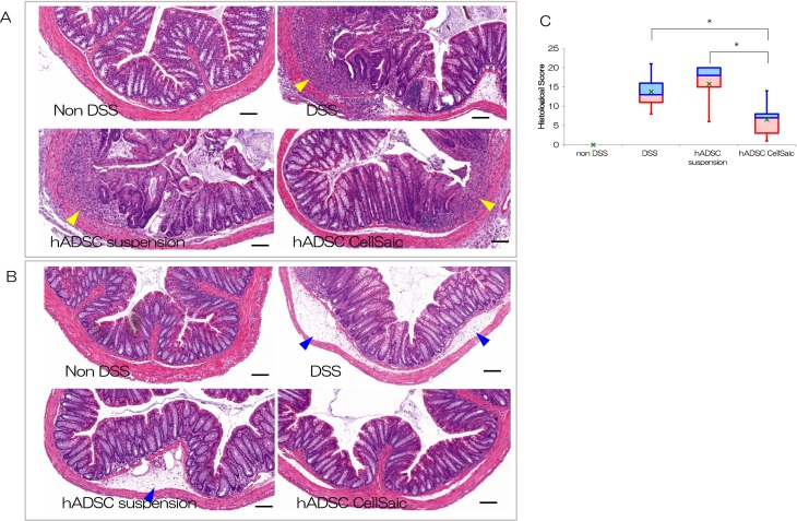
Fig. 3.
ADSC CellSaic decreased inflammatory cells and repaired ulceration in histological analysis. Histopathological comparison of colitis in lower colon at day 14. There was no ulceration and no infiltration in the non-DSS group. In the DSS group and hADSC suspension group, ulceration and infiltration are more severe than in the hADSC CellSaic group. The arrows indicate ulceration and infiltration. Scale Bar is 100 µm. (A) Histopathological comparison of colitis in the rectum at day 14. There was no edema in the non-DSS group. In the DSS group and hADSC suspension group, edema was more severe than in the hADSC CellSaic group. The arrows indicate edema. Scale bar is 100 µm. (B) Histological evaluation of the colon at day 14. The value of 75% – median (blue box), median – the value of 25% (red box), the value of average (green cross). Non-DSS group [no treatment with DSS, n = 5], DSS group [no cell transplantation into DSS mice, n = 5], hADSC suspension i.p. group [1.2 × 10⁶ hADSCs in suspension intraperitoneally administered to DSS mice, n = 5], hADSC CellSaic i.p. group [CellSaics of 1.2 × 10⁶ hADSCs and 1 mg u-pieces, intraperitoneally administered to DSS mice, n = 5]. *p < 0.05. hADSC CellSaic group compared with DSS group and hADSC suspension group.

