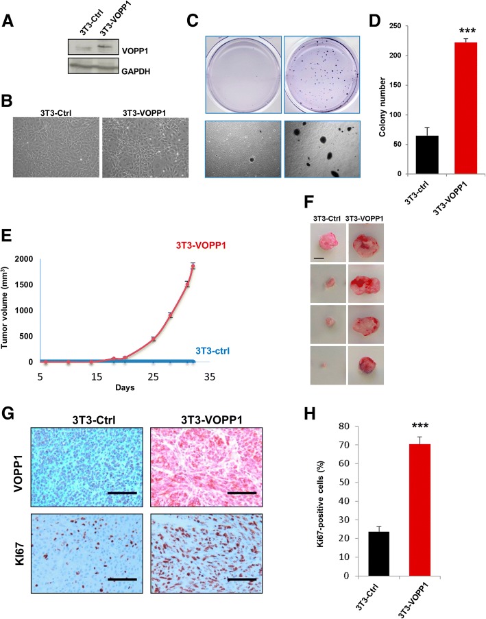
Fig. 5.
VOPP1 induces cell transformation in vitro and tumorigenesis in vivo. a NIH3T3 cells were stably transfected with control VOPP1-expressing vectors. Cellular extracts were immunoblotted with anti-VOPP1 and anti-GAPDH (loading control) antibodies. b Morphology of NIH3T3 cells stably expressing VOPP1 (3T3-VOPP1) or empty vector (3T3-ctrl). c Low-magnification photographs represent the results of soft agar colony formation assays showing the number and size of colonies formed by 21 days after plating 3T3-Ctrl and 3T3-VOPP1 cells. d The quantitative results of colony numbers are expressed as mean ± SEM of a representative experiment (n = 4). e 2.106 3T3-Ctrl and 3T3-VOPP1 cells were subcutaneously injected into the flanks of SCID mice, and tumor growth was measured over time (n = 10 per each group) up to 35 days. f 3T3-Ctrl and 3T3-VOPP1 mice were sacrificed when tumors reached a volume of approximately 2 cm3; otherwise, they were sacrificed at the end of the experiment (week 25, for 3T3-VOPP1 mice only), representative images of tumors (scale bar, 1 cm). g Immunohistochemical staining of 3T3-Ctrl and 3T3-VOPP1 tumors with indicated antibodies (scale bar = 100 μm). h Quantification of Ki67+ proliferating cells in tumors. Statistical analysis was made by performing the Student t test (*p < 0.05; **p < 0.01; ***p < 0.001)

