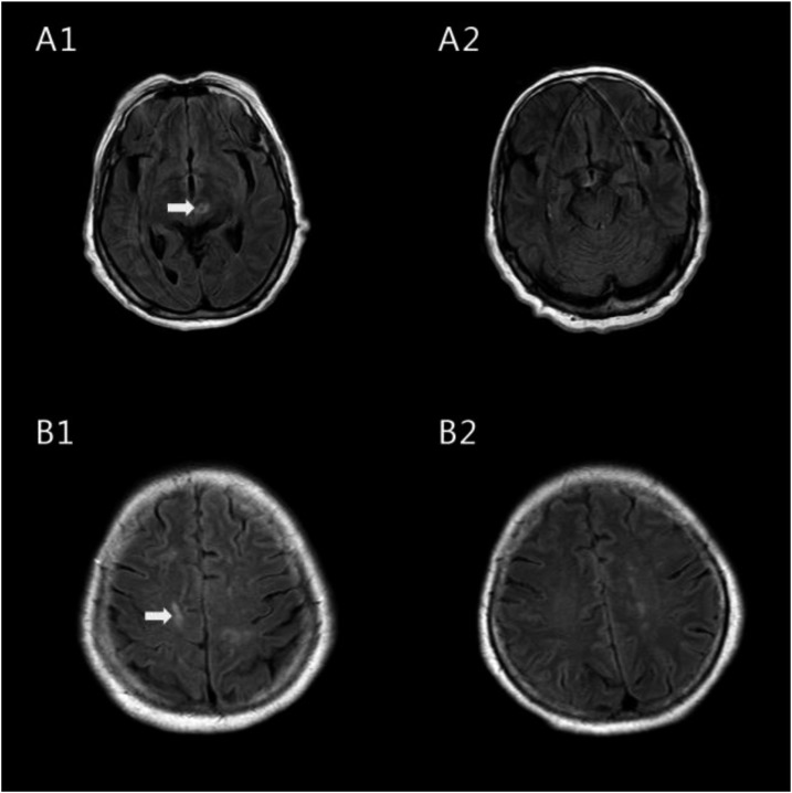
Figure 1.
Flow alternating inversion recovery-magnetic resonance imaging of the female patient’s brain before and after treatment. The arrows indicate the hyperintense signal that suggests multiple inflammatory foci in specific areas before treatment (A1, B1). They disappeared after treatment (A2, B2).

