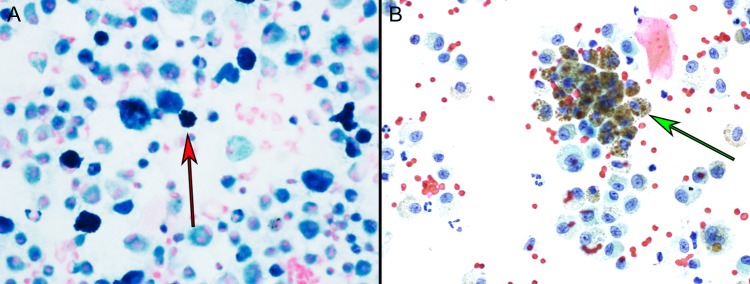
Figure 2:
Hemosiderin-laden macrophages seen on bronchoalveolar lavage smears with (A) Prussian blue iron stain in which the hemosiderin stains a dark blue color inside the macrophages (red arrow) and (B) alcohol Papanicolaou stain in which the hemosiderin stains a brown color in the macrophages (green arrow). Each image is at ×40 magnification.

