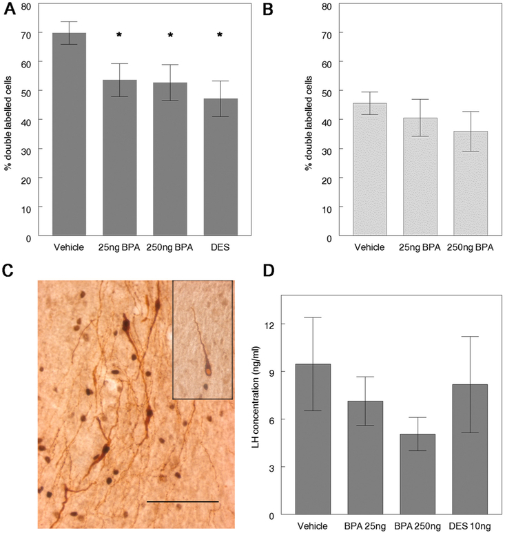
Fig. 2.
Females exposed perinatally to BPA revealed evidence of decreased activation of GnRH neurons in conjunction with a steroid-induced LH surge. Perinatal BPA exposure decreased activation of GnRH neurons. [A] The percent of double labeled GnRH neurons (GnRH+cFos) were significantly reduced in BPA and DES–exposed females relative to controls at 3–6 months of age (Kruskal Wallis: p=0.009); * p < 0.05 by Mann Whitney U compared to vehicle treated animals, n = 11–16/treatment. [B] By 9–12 months of age, the percent of double-labeled GnRH neurons in BPA exposed females did not differ from controls, n = 6-8/treatment. [C] Double-labeled GnRH neurons are characterized by brown cytoplasm (GnRH immunoreactivity) and black nuclei, denoting the presence of cFos protein (box inset: single-labeled GnRH neuron characterized by brown cytoplasm and a pale nucleus). Scale bar=100 μm. [D] No significant differences in mean serum LH concentrations were observed in relation to treatment; however, LH levels in 250 ng BPA females were reduced in 3–6 month old animals relative to controls, n = 11-16/treatment.

