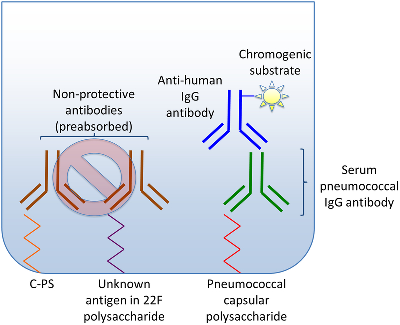
Figure 1. WHO ELISA for measurement of pneumococcal IgG antibodies.
The figure represents a typical plate well in WHO ELISA. The left side of the figure depicts the initial pre-absorption of sera with cell wall polysaccharide (C-PS) and 22F polysaccharide to neutralize non-protective antibodies (shown in brown).
The right side of the figure illustrates binding of serum pneumococcal IgG antibodies (shown in green) to serotype-specific pneumococcal capsular polysaccharide. Anti-human IgG antibodies (shown in blue) are bound to serum IgG antibodies. Using a chromogenic substrate, optical density is measured, and then converted to antibody concentration.

