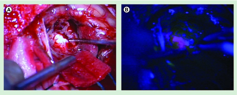
Figure 2. Intraoperative image after complete removal of a cerebellar breast cancer metastasis.
5-aminolevulinic acid was administered to the patient 5 h prior to surgery. (A) Picture taken with the help of the operation microscope under white-light illumination. Suction device and pointer of neuronavigation are visible. (B) Picture taken with the help of the operation microscope under 400-nm blue-light illumination. Suction device and bipolar forceps are visible. A positive protoporphyrin IX staining of the tumor-surrounding tissue can be noticed. The tissue proved to be tumor negative in subsequent histological analysis.

