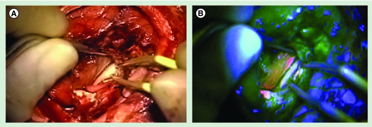
Figure 3. Ependymoma WHO grade III.
(A) Intraoperative view into the ventricle of the left hemisphere of the brain of a patient with ependymoma WHO grade III under white-light. Identical view through the operation microscope with switched-on blue excitation light. (B) The fluorescent surface of the ependymoma is clearly visible and easy to distinguish from the surrounding brain and ventricle's ependyma.

