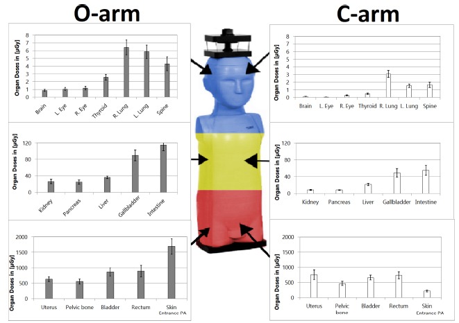
Fig. 2.

Absorbed doses comparison for each organ between one 3D acquisition with the O-arm and one minute of fluoroscopy with the C-arm. Direct irradiation on the pelvic organs is coloured by red. Scattered irradiation on abdominal organs is coloured yellow and on thoracic and upper organs blue, with decreasing level respectively (R., right; L., left; PA, posteroanterior).
