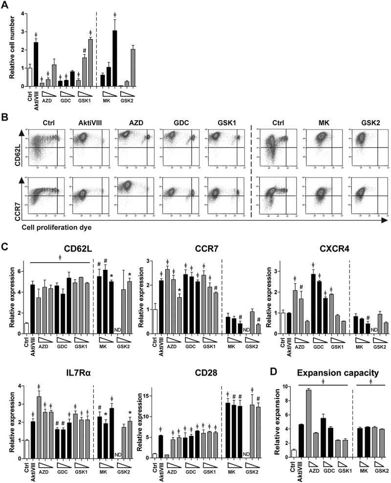
Figure 2.

Allogeneic stimulated AKT-inhibited CD8+ T cells retain an early memory phenotype and superior expansion capacity. CD8+ TN cells were stimulated with allogeneic mDCs in presence of DMSO (Ctrl) or AktiVIII (12µM), AZD (12–8-4 µM), GDC (40–30–20 µM), GSK1 (7.5–5-2.5 µM), MK (10–5-2.5 µM) and GSK2 (30-20-10 µM). MK and GSK2 were evaluated in different donors. (A) Relative cell number of AKT-inhibited CD8+ T cells compared to Ctrl T cells. (B) Representative plots of CD62L and CCR7 expression during proliferation of Ctrl, 12 µM AktiVIII, 8 µM AZD, 20 µM GDC, 5 µM GSK1, 5 µM MK and 20 µM GSK2-treated T cells, gated on viable CD8+ T cells. (C) Relative expression of CD62L, CCR7, CXCR4, IL7Rα and CD28 within proliferated CD8+ T cells. (D) Relative expansion of AktiVIII (12 µM), AZD (12–8 µM), GDC (40–30 µM), GSK1 (7.5–5 µM), GSK2 (20-10 µM) or MK (10-5 µM) treated cells during re-stimulation with allo-mDCs for 7 days after removal of AKT-inhibitor. Mean + SD of one representative donor out of 2–5 donors, n = 3 per experiment. Statistical analysis was performed using One-way ANOVA followed by Bonferroni’s Multiple Comparison Test of AKT-inhibited versus Ctrl T cells within donors, ǂp < 0.001, #p < 0.01, *p < 0.05.
