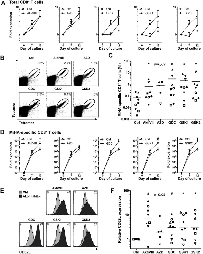
Figure 5.

AKT-inhibition preserves CD62L expression while promoting expansion of antigen-specific CD8+ T cells. CD8+ T cells were stimulated with peptide-loaded mDCs for 12 days in the absence (Ctrl) or presence of AktiVIII (12 µM), AZD (4 µM), GDC (10 µM), GSK1 (2.5 µM), or GSK2 (10 µM). (A) Expansion of total CD8+ T cells. (B) Representative tetramer staining and (C) percentages of MiHA-specific CD8+ T cells on day 12 of culture. (D) Expansion of MiHA-specific CD8+ T cells calculated from an estimated precursor frequency of 1:107. (E) Overlays of representative CD62L staining gated on Ctrl or AKT-inhibited MiHA-specific CD8+ T cells. Numbers represent Median Fluorescence Intensity of CD62L. (F) Relative CD62L expression of AKT-inhibited compared to Ctrl MiHA-specific CD8+ T cells on day 12 of culture. (C&F) Individual donors are depicted with unique symbols. Statistical analysis was performed using (A&D) Two-way ANOVA followed by a Bonferroni post-hoc test or a (C&F) one-tailed Wilcoxon matched-pairs signed rank test. ǂp < 0.001, #p < 0.01, *p < 0.05 compared to matched Ctrl cultures.
