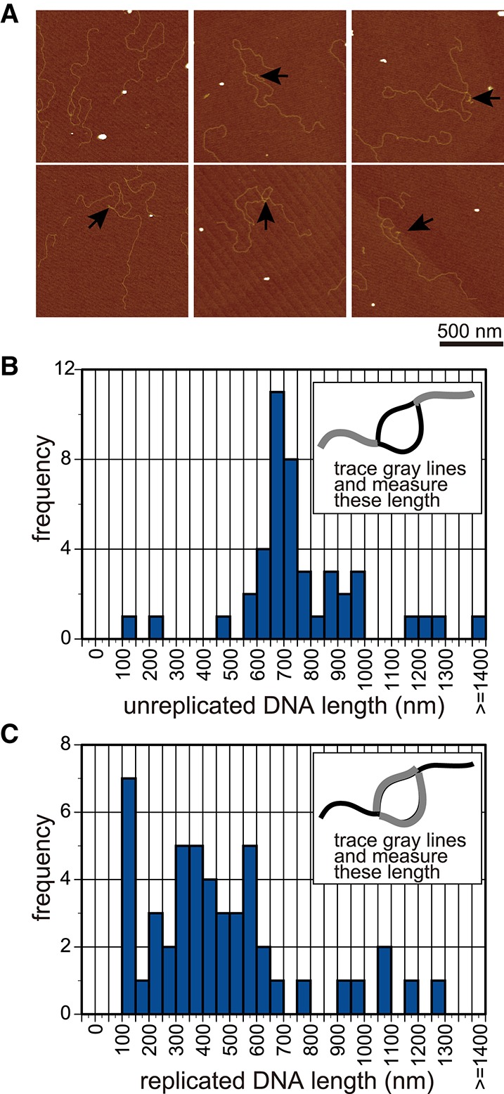
Figure 2.

AFM analysis of the replication pausing. (A) AFM images of the DNA that was partially replicated. DNA was purified from the reaction of lane 6 in Figure 1C via treatment with phenol, cleaved with the restriction enzyme ScaI at the opposite side of the origin (Supplemental Fig. S2A,B), and observed by AFM. The ssDNA region, which was unwound and might not have been used for the synthesis of a complementary strand, was observed as the shrinking structure indicated by arrows. (B,C) The lengths of the unreplicated DNA (B) or replicated DNA (C) were measured and are shown in the histogram (n = 44 and n = 48, respectively).
