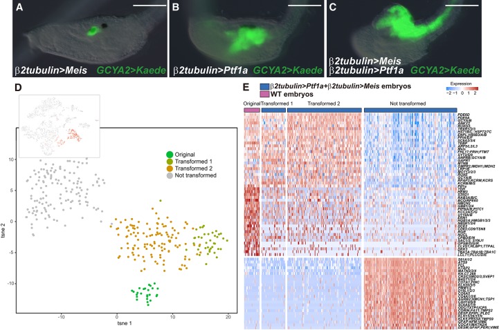
Figure 3.
A Ptf1a + Meis cocktail promotes the differentiation of DA neurons/coronet cells. (A–C) Head regions of larvae that were injected with a GCYA2>Kaede reporter gene that is specifically expressed in DA neurons/coronet cells (see Supplemental Fig. S2). (A) The embryo was coinjected with a β2tubulin>Meis transgene. Misexpression of Meis does not alter the normal expression of the reporter gene within DA neurons/coronet cells (100 of 100 larvae displayed this expression pattern). (B) The embryo was coinjected with the β2tubulin>Ptf1a transgene. Kaede expression is expanded into posterior regions of the sensory vesicle and anterior neural tube (104 of 104 larvae displayed this expression pattern) (see Fig. 2C). (C) The embryo was coinjected with both β2tubulin>Ptf1a and β2tubulin>Meis transgenes. The GCYA2 reporter gene is now expressed throughout the entire CNS (89 of 89 larvae displayed this expression pattern). Bar, 100 µm. (D, top) A tSNE projection map of late tail bud stage embryos expressing both the Ptf1a and Meis transgenes. Red dots correspond to cells expressing the β2tubulin>CFP reporter gene, which identifies cells that misexpress Ptf1a and Meis. (Bottom) tSNE subclustering of coexpressing cells. (E) Heat map of native DA neurons/coronet cells, transformed cluster 1, transformed cluster 2, and untransformed cells. A select group of genes encoding cellular effectors and TFs is shown. Clusters 1 and 2 display transcriptome profiles that are very similar to those seen for native coronet cells. The untransformed cells are likely to correspond to epidermis based on their transcriptome profiles.

