Figure 4.
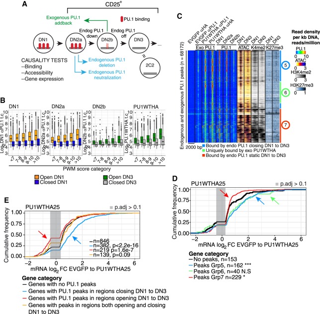
Causality test: sites bound by exogenous PU.1 that mediate function in pro-T cells. (A) Schematic of experiments shown in this figure and Supplemental Figures S4 and S5: acute gain and loss of function of PU.1 in developing primary pro-T cells. CD25+ cells (DN2 to DN3) are cells within the T-cell pathway. (B) Comparison of site occupancies by endogenous PU.1 (DN1-DN2b, detected by αPU.1) and by exogenous PU.1 (PU1WTHA, detected by αHA) as a function of PWM score, at “open” and “closed” sites. (C) Heatmap of binding of exogenous PU1WTHA in CD25+ DN2b/DN3 primary cells (Exo PU.1) compared with endogenous PU.1 in DN1, DN2a, and DN2b, ATAC-seq in DN1 and DN3, and H3K4me2 and H3K27me3 in DN1 and DN3 cells. Color scales: tag count densities. (D) Association of Groups 5, 6, and 7 sites with changes in linked gene expression induced by exogenous PU1WTHA in CD25+ cells. (***) Kolmogorov–Smirnov P-value ≤0.0001; (*) P-value ≤0.05. (E) Changes in gene expression induced by acute PU1WTHA introduction in genes linked to sites that would normally close (blue) or open (red) from DN1 to DN3 as compared to genes with no PU1WTHA peaks. Genes with a mixture of opening and closing peaks are also shown. Genes are those with significant differential expression induced in cells remaining CD25+.

