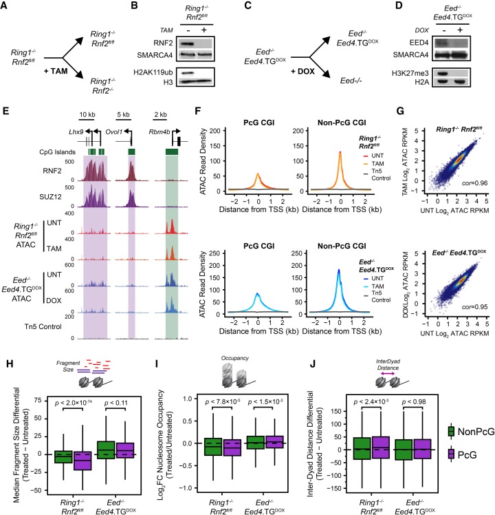
Figure 3.
PRC1 contributes toward nucleosome spacing and occupancy but not chromatin accessibility. (A) A schematic depicting the treatment of Ring1−/−;Rnf2fl/fl ESCs with 4-hydroxytamoxifen (TAM) to generate PRC1-null ESCs. (B) A Western blot analysis of untreated and TAM-treated Ring1−/−;Rnf2fl/fl ESCs for RNF2 and H2AK119ub1. (C) A schematic depicting the treatment of Eed−/−;Eed4.TGDOX ESCs with doxycycline (DOX) to generate PRC2-null ESCs. (D) A Western blot analysis of untreated and DOX-treated Eed−/−;Eed4.TGDOX ESCs for EED and H3K27me3. (E) Genome screenshots of chromatin accessibility, as measured by ATAC-seq, at two PcG-occupied promoters (purple boxes) and one PcG-free promoter (green box) before and after conditional deletion of PRC1 or PRC2 from mouse ESCS. (F) A metaplot analysis for Ring1−/−;Rnf2fl/fl and Eed−/−;Eed4.TGDOX ATAC-seq before and after TAM or DOX treatment, respectively, at PcG-occupied (n = 4020) or non-PcG CGI promoters (n = 10,251), centered on TSSs. (G) A scatterplot analysis comparing untreated and treated reads per kilobase per million (RPKM) for Ring1−/−;Rnf2fl/fl and Eed−/−;Eed4.TGDOX ATAC-seq at all CGI promoters. (H) A box plot comparing the change in median Tn5-tagmented fragment sizes for PcG and non-PcG CGI promoters in Ring1−/−;Rnf2fl/fl and Eed−/−;Eed4.TGDOX ATAC-seq data sets before and after TAM or DOX treatment, respectively. A decrease in median fragment size reflects a shift toward a more nucleosome-free state. (I) A box plot quantifying the log2 fold change (log2FC) in NucleoATAC-derived nucleosome occupancy for PcG and non-PcG CGI promoters in Ring1−/−;Rnf2fl/fl and Eed−/−;Eed4.TGDOX ATAC-seq data sets before and after TAM or DOX treatment, respectively. (J) A box plot comparing the difference in median inter-dyad distances for PcG and non-PcG CGI promoters in Ring1−/−;Rnf2fl/fl and Eed−/−;Eed4.TGDOX ATAC-seq data sets before and after TAM or DOX treatment, respectively.

