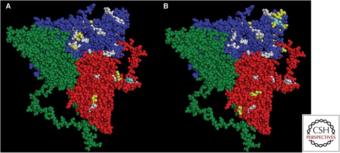
Figure 4.
Hepatitis A virus (HAV) protomer (Wang et al. 2015) showing residues encoded by rare codons (white), and residues replaced in VP3 and VP1 in quasispecies placed under the immune pressure of monoclonal antibodies (mAbs) (yellow). Although residues undergoing replacement are located very close to residues encoded by rare codons, there is a general lack of coincidence with only few matching positions (clear blue). (A) Selection with H7C27 mAb. (B) Selection with K34C8 mAb.

