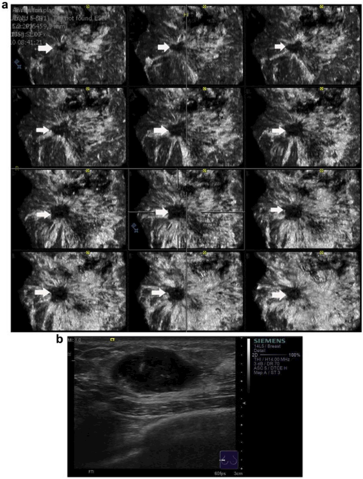
Figure 10.
(a) Automated breast volume scanner images of an invasive ductal carcinoma of the breast in a multislice view from the skin down to the thoracic wall in the coronal plane (the slices are 0.5 mm). (b) Handheld B- mode ultrasound. The typical retraction phenomenon of the mass is observed in several, consecutive coronal planes (arrowhead on the right).This indicates the conditions of the masses at different depths. Reprinted from (Chen, et al. 2013).

