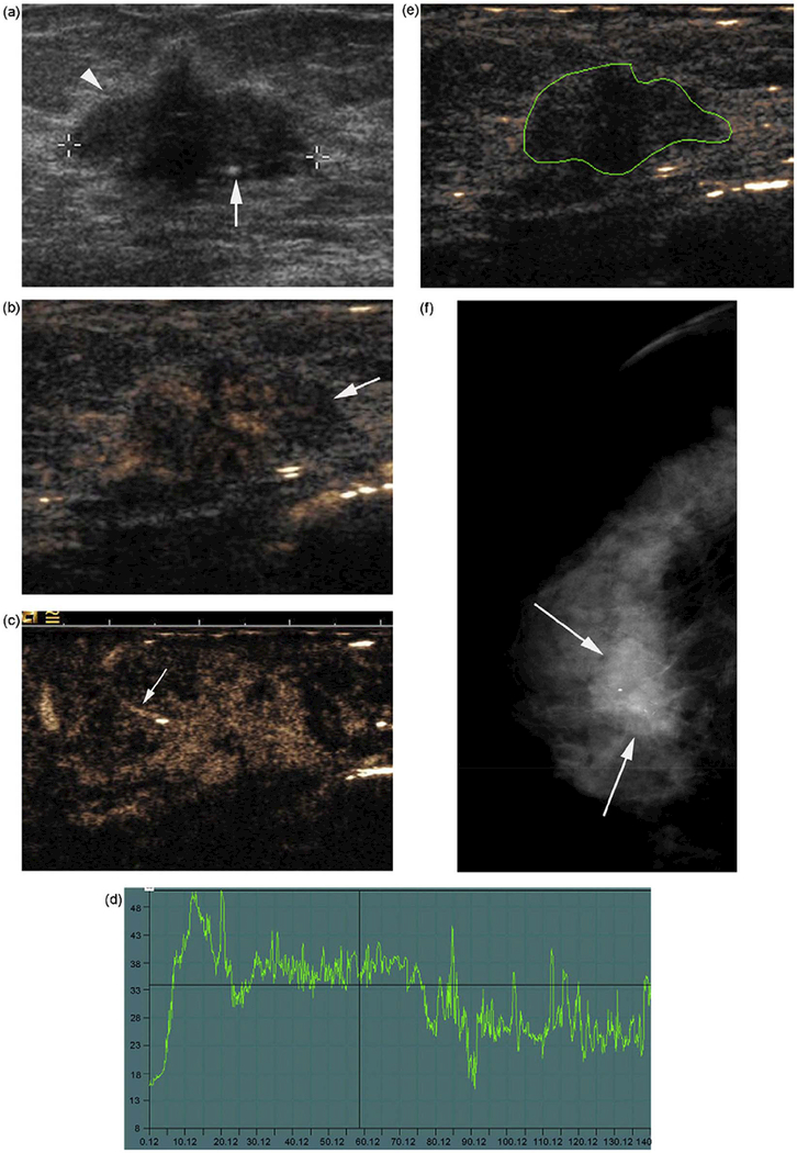
Figure 5.
Contrast US before and after contrast medium injection in a 66-year-old woman with 23–mm, ductal, infiltrative carcinoma. (a) B mode sonography. (b) In the contrast mode (SonoVue ®) with Coherence Pulse Sequencing and B mode, the tumor is strongly enhanced after injection. Vessels are located in the peripheral area of the lesion. (c) In the contrast mode with Coherence Pulse Sequencing only, the tumoral-feeding artery is visible outside of the lesion (arrows). (d) Dynamic curve of enhancement after injection. Enhancement is fast, i.e. the delay of peak enhancement = 10 s, and with a wash-out phase (total time: two min). (e) Region of interest (ROI) on the tumor to obtain enhancing curves. (f) Mammogram. Cranio-caudal view of the right breast. Reprinted from (Balleyguier, et al. 2009)

