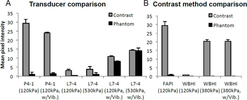
Figure 5.

Comparison of contrast image acquired with A) transducers of differing center frequency, and B) differing contrast imaging methods. A) Contrast and phantom signal amplitude captured with FAPI using P4-1 (1.8MHz) and L7-4 (3.9MHz), with and without vibration. L7-4 Tx frequency was chosen based upon its spectral efficiency (figure 2), such that the transducer would be comparably sensitive to both the fundamental and 2nd harmonic frequencies. B) Comparing FAPI with wide beam harmonic imaging (WBHI), the Verasonics’ line-by-line contrast imaging method using pulse inversion.
