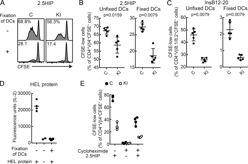
Figure 10.
Reduced presentation of extracellular peptides by DCs from knock-in mice. (A–C) Presentation of soluble 2.5HIP peptide (A and B) or InsB12-20 peptide (C) by DCs fixed with paraformaldehyde. CFSE-labeled CD4 T cells from BDC-2.5 Tg (A and B) or 8F10 Tg mice (C) were co-cultured with fixed or unfixed DCs from M98A/M98A (KI) or control (C) mice. On day 3, CD4 cells were analyzed for CFSE dilution. (A) Representative flow-cytometric analysis of CFSE dilution by T cells. (B) Summary plots of data from A. n = 5 samples/group in B and C. (D) Presentation of HEL protein by DCs fixed with paraformaldehyde. The 21.30 hybridoma was cultured with fixed or unfixed DCs from control mice. On day 1, IL-2 released into the supernatant was measured by ELISA (n = 2–4 samples/group). (E) Presentation of 2.5HIP peptide by DCs treated with cycloheximide. CFSE-labeled CD4 T cells from BDC-2.5 Tg mice were co-cultured with cycloheximide-treated or untreated DCs from M98A/M98A (KI) or control (C) mice. On day 3, CD4 cells were analyzed for CFSE dilution (n = 4 samples/group). Data representative of two independent experiments. Statistical analyses were performed with the Mann-Whitney test (B and C); mean (D) or mean ± SD (B, C, and E) are shown.

