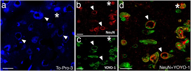
Figure 2.
Donut-like staining of neurons in poorly perfused sections was also observed with other nuclear markers. Frozen sections exhibited abnormal (donut-like) staining pattern with nuclear stains TO-PRO-3 (triangles in a) similar to those seen with NeuN (triangles in b); and YOYO-1 (triangles in c). Merged image (d) illustrates the colocalization of abnormal nuclear staining with YOYO-1 and NeuN (triangles in d). Intact cells are shown by asterisks. Scale bars: 20 µm.

