Figure 4.
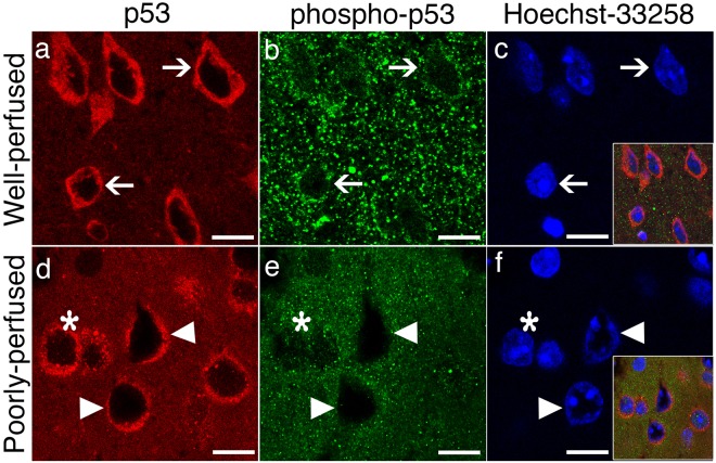
Poor perfusion instantly activated p53-mediated death signaling. In well-perfused brains (upper row), p53 immunolabeling (red) exhibits a predominantly cytoplasmic and diffuse staining (arrows, a), whereas anti-serine15-phospho-p53 antibodies weakly immunostained the nuclei (arrows, green, b). (c) Illustrates the intact nuclei identified by Hoechst staining (arrows, blue). The inset is the merged image of (a,b,c). In poorly perfused brain sections (lower row), p53 is still predominantly expressed in the cytoplasm but assumed a granular pattern (red, d). Triangles point at the swollen donut-like stained nuclei (d–f). These nuclei were not immunolabeled with phospho-p53 antibodies (green, e), whereas other intact nuclei preserved phospho-p53 immunopositivity (*). Coarse granular cytoplasmic phospho-p53 immunostaining (b) was replaced by a fine granular pattern in poorly perfused brain sections (e,f) illustrates the nuclei identified by Hoechst staining (blue). The inset is the merged image of (d,e and f). Scale bars: 10 µm.

