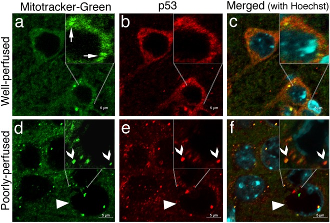
Figure 5.
p53 translocates to mitochondria in poorly perfused brain sections. In well-perfused brains (upper row), p53 immunolabeling (red) exhibits a predominantly diffuse cytoplasmic staining (b) and occasional colocalization with mitochondria visualized with mitotracker-green (arrows, green, a). (c) illustrates the nuclei identified by Hoechst staining (blue) on a merged image of (a and b). In poorly perfused brain sections (lower row), p53 immunopositive granules (arrowheads, red, e) sharply colocalize with mitochondria (arrowheads, green, d). (f) Illustrates the nuclei identified by Hoechst staining (blue) on the merged image of (d and e). Triangles point at a swollen donut-like stained nucleus. Insets show magnified images. Images were taken at 0.2 µm thickness by laser scanning confocal microscope. Scale bars: 5 µm.

