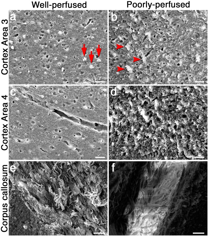
Figure 7.
Representative SEM images taken from cortex 3, 4 and corpus callosum. A well-perfused section from cortex 3 displays relatively round neuron somas (arrows in a), whereas dysmorphic and swollen neural somas are seen in poorly-perfused samples (arrowheads in b). Integrity of the intercellular tissue is also disrupted in poorly-perfused sections (b). Fine structure of blood vessels in cortex 4 is clearly discernable in well-perfused samples (c) unlike poorly-perfused samples (d). Neuronal fibers are well-preserved in well-perfused sections taken from corpus callosum (e), whereas intense electron discharges emerge as a result of poor perfusion (f). Scale bars: 20 µm.

