Figure 3.
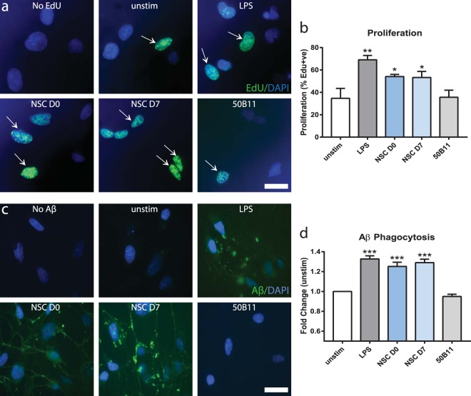
NSCs modulate microglial function in vitro: a possible therapeutic mechanism. Representative immunocytochemistry images and quantification of in vitro microglial proliferation (a,b) and phagocytosis of Aβ (c,d) with and without co-cultured NSCs. Proliferating EdU+ microglia (green) were increased by addition of LPS and co-culture with undifferentiated and differentiated NSCs vs. unstimulated and 50B11 controls (a). Quantified percentage proliferation was significantly increased in NSC co-cultures vs. the control groups (b; p < 0.05; ANOVA). NSCs also enhanced phagocytic activity of microglia (c), evidenced by increased accumulation of fluorescently-labeled Aβ vs. unstimulated and 50B11 controls. Quantified phagocytosed fluorescence was significantly increased by addition of LPS and NSC co-cultures vs. unstimulated and 50B11 controls (d; p < 0.0001; ANOVA). Data are representative images or mean ± SEM (*p < 0.05, ***p < 0.0001) of 3–5 independent experiments (n = 3–6 per condition). Scale bar 50 µm. Abbreviations: unstim, unstimulated; D0, day 0 undifferentiated NSCs; D7, day 7 differentiated NSCs; EdU, 5-ethynyl-2′-deoxyuridine.

