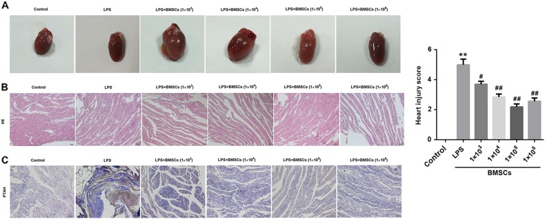
Fig. 2. Effect of BMSCs pre-treatment on hearts in LPS-induced DIC model rats.
Six hours after LPS injection, the rats were killed and their heart tissue sections were examined by hematoxylin and eosin (HE) and Mallory’s phosphotungstic acid hematoxylin (PTAH) staining. a Representative images of heart tissues. b Representative images of HE staining of heart tissues from rats. c Representative images of PTAH staining of heart tissues from rats. Fibrin was red-stained in HE staining and violet blue-stained in PTAH staining. The bar graphs show the heart injury score. N = 10/group.*P < 0.05, vs. control; #P < 0.05, ##P < 0.01, vs. LPS

