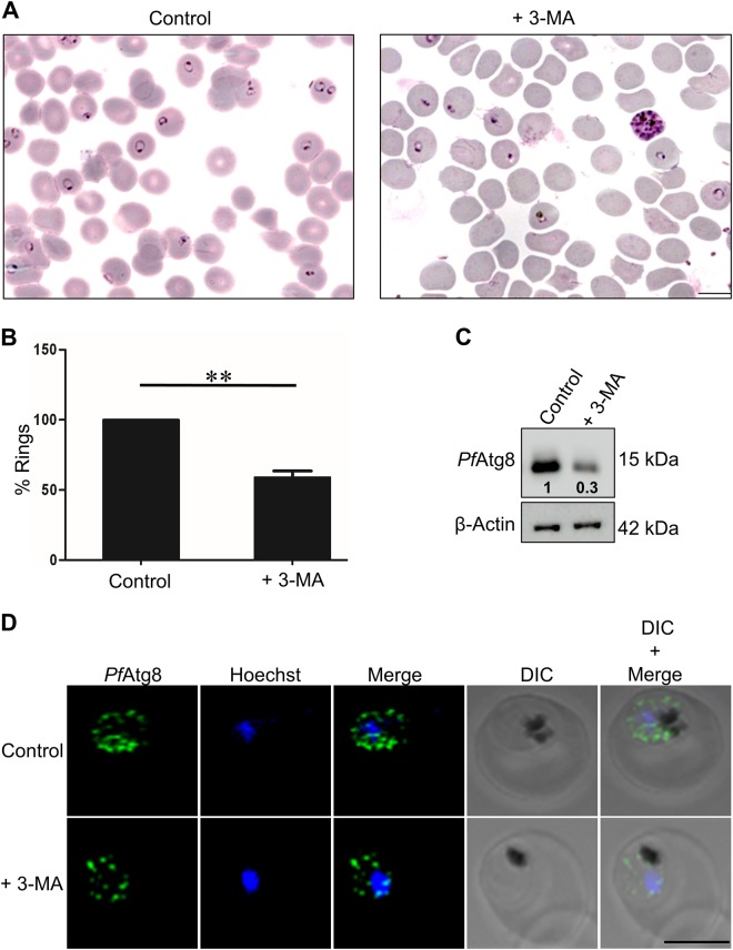
Fig. 1. Inhibition of basal autophagy results in reduced reinvasion and PfAtg8 expression.
a Giemsa stained smears showing rings in control and 3-MA treated parasites. Highly synchronized parasites at late schizonts stage (38 h.p.i) were incubated in complete medium (control) with or without 3-MA (5 mM) for 2 h, and invasion was monitored by counting number of rings in the next cycle. Scale bar: 10 µm. b Graph representing percent rings in control and 3-MA treated parasites. The data presented are mean of 5 individual experiments. Error bars show the standard deviation. **P < 0.05. c Immunoblot analysis of PfAtg8 expression levels. Lysates were prepared from tightly synchronized parasites at trophozoite stage incubated in complete medium with or without 3-MA for 2 h. Immunoblot was probed using custom generated anti-PfAtg8 antibodies.β-actin was probed as loading control. Number below the bands indicate fold difference as compared to the normalized control. Uncropped blots are shown in Supplementary Figure 5. d PfAtg8 immunofluorescence staining in control and 3-MA treated parasites. Hoechst was used as a DNA marker. Scale bar: 5 µm

