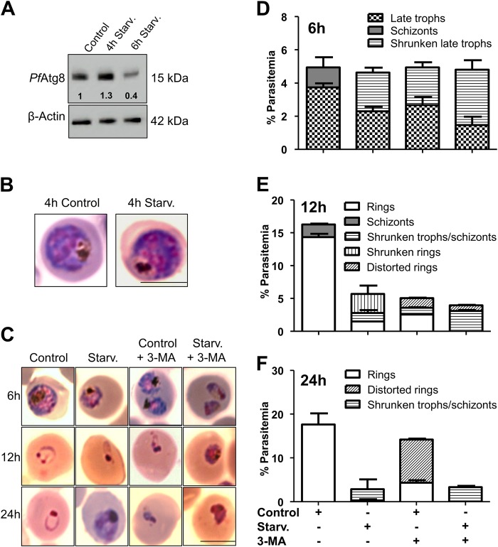
Fig. 3. Duration of starvation-induced autophagy determines parasite survival.
a Immunoblot analysis of PfAtg8 expression levels upon 4 and 6 h starvation. Lysates from trophozoite stage parasites cultured in complete or starvation medium for 4 and 6 h were analyzed using immunoblotting by probing with custom generated anti-PfAtg8 antibodies. Uncropped blots are shown in Supplementary Figure 6. b Morphological features of control and 4 h starved parasites stained with Giemsa. Scale bar: 5 µm. c Prolonged starvation induces abnormal morphology. Parasites at late trophozoite stage were incubated in control, control + 3-MA, starvation or starvation + 3-MA media for 6, 12 and 24 h. Parasite morphology was assessed in Giemsa stained smears. Scale bar:5 µm. d–f Graph representing parasites with starvation-induced morphological changes after 6, 12 and 24 h incubation in control, control + 3-MA, starvation or starvation + 3-MA media. Data represented is mean of 5 individual experiments. Number of parasites scored for morphology, n = 2500. Error bars show standard deviation

