Figure 5.
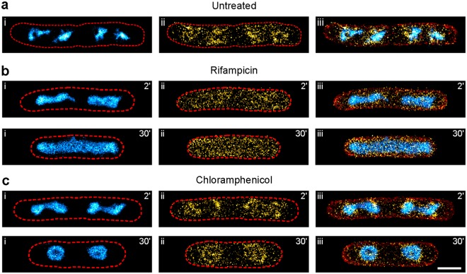
Combination of PALM and PAINT for parallel 3-colour SMLM imaging. Strain KF26 was grown in LB medium at 32 °C, chemically fixed and imaged (see Methods section). Shown are the nucleoid (i, cyan hot), the RNA polymerase β′ subunit (ii, PAmCherry1 fusion, yellow hot) and the overlay (iii) including the membrane PAINT image (red). (a) Representative 3-colour image of untreated KF26 cells. Cell outlines (red dashed lines) were determined using the Potomac Red membrane PAINT image. (b) Representative images of KF26 cells treated with 100 µg/ml rifampicin for the indicated duration. (c) Representative images of KF26 cells treated with 50 µg/ml chloramphenicol for the indicated duration (scale bar: 1 µm).

