Figure 5.
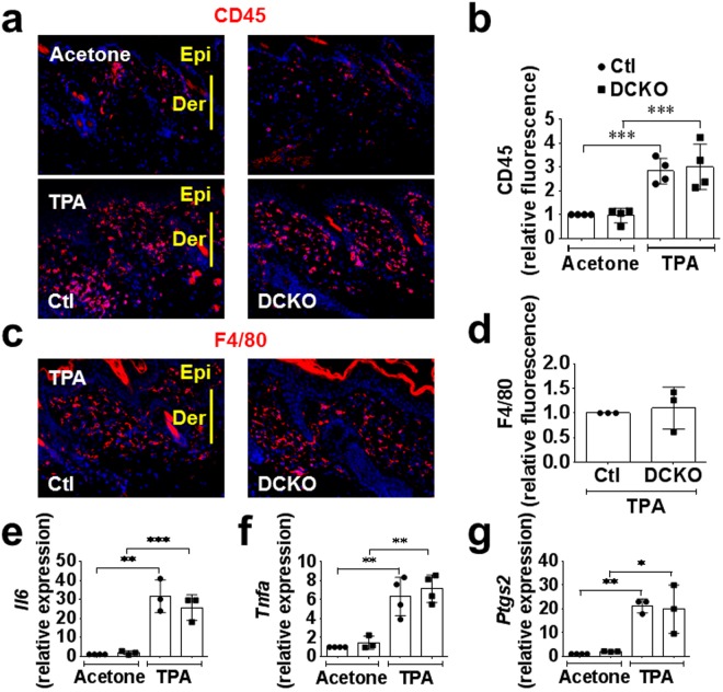
Lymphocyte or macrophage infiltration into the skin in response to TPA was unaffected by Vegfr2 and endothelial Fgfr1/2 deficiency. (a,b) Immunofluorescence staining (a) and quantitation (b) showing increased CD45-positive inflammatory cells seven days post TPA challenge compared to vehicle (acetone). No difference was observed in both DFF control (Ctl) and DCKO responses. (c,d) Immunofluorescence staining (c) and quantitation (d) showing increased F4/80-macrophages seven days post TPA challenge. No difference was observed in control and DCKO mice. (e,f) Increased inflammatory cells markers, Tnfa, Il6 and Ptgs2, seven days post TPA challenge in both control and DCKO mice compared to vehicle treated mice. No difference was observed between genotypes. For quantitative data, each symbol represents one mouse. Mann-Whitney U test was used to analyze significance (*p < 0.05; **p < 0.01; ***p < 0.001) compared to vehicle (Acetone). Ear skin (a,c) sections were imaged with a 10X objective.

