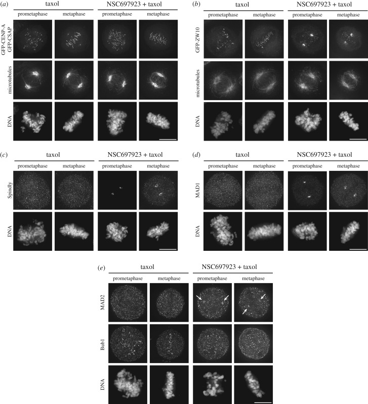
Figure 3.
NSC697923 induces accumulation of a subset of dynein interactors at the mitotic spindle poles. (a) Representative immunofluorescence images of stably expressed GFP-CENP-A and GFP-CSAP, microtubules (DM1A) and DNA (Hoechst) after treating the cells for 15 min with the indicated compounds. Scale bar, 10 µm. (b) Representative immunofluorescence images of stably expressed GFP-ZW10, microtubules (DM1A) and DNA (Hoechst) after treating the cells for 15 min with the indicated compounds. Scale bar, 10 µm. (c) Representative immunofluorescence images of Spindly and DNA (Hoechst) after treating the cells for 15 min with the indicated compounds. Scale bar, 10 µm. (d) Representative immunofluorescence images of MAD1 and DNA (Hoechst) after treating the cells for 15 min with the indicated compounds. Scale bar, 10 µm. (e) Representative immunofluorescence images of MAD2, Bub1 and DNA (Hoechst) after treating the cells for 15 min with the indicated compounds. Arrows indicate weak, but detectable localization of MAD2 at the spindle poles. Scale bar, 10 µm.

