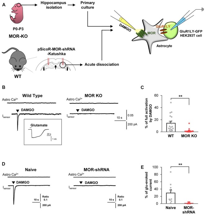
Figure 2.
DAMGO-induced astrocytic glutamate release is caused by MOR activation. (A) Schematic diagram for sniffer-patch with primary cultured hippocampal astrocytes prepared from MOR knockout (KO) mouse or acutely dissociated astrocytes from pSicoR-MOR-shRNA-Katushka-injected hippocampus of wild-type (WT) mouse. (B) Representative traces of Ca2+ response and inward current induced by DAMGO in primary cultured hippocampal astrocyte of WT and MOR KO mice. Inset indicates an example trace of full activation current induced by bath application of 1 mM glutamate. (C) Summary bar graph for DAMGO-induced glutamate current normalized by the full activation current in WT and MOR KO mice. Numbers in the bar graph indicate the number of cells tested from at least three independent mice for each group. Unpaired t-test (**P < 0.01). (D) Representative traces of Ca2+ response and inward current naïve or MOR-shRNA-infected astrocyte. (E) Summary bar graph for DAMGO-induced glutamate current normalized by the full activation current in naive and MOR-shRNA-infected astrocytes. Numbers of tested cells are indicated on each bar. Unpaired t-test with Welch’s correction (**P < 0.01).

