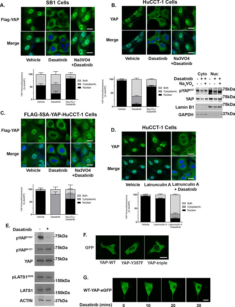
Figure 4. Phosphorylation of YAPY357 regulates nuclear retention in cholangiocarcnoma.
(A) Representative images of FLAG immunofluorescence in the SB1 CCA cell line incubated with vehicle, dasatinib (100 nM, 1 hour), or dasatinib (100 nM, 1 hour) and sodium orthovanadate (10 mM, 1 hour) (upper panel) and quantification of YAP nuclear localization (lower panel). (B) Representative images of YAP immunofluorescence in HuCCT-1 cells incubated with vehicle, dasatinib (100 nM, 1 hour), or dasatinib (100 nM, 1 hour) and sodium orthovanadate (10 mM, 1 hour) (upper panel), quantification of YAP nuclear localization (lower left panel), and cell fractionation evaluated by immunoblot (lower right panel). Lamin B1 and GAPDH were used as nuclear and cytoplasmic markers respectively. (C) Representative images of FLAG immunofluorescence in HuCCT-1 cells transfected with FLAG-5SA-YAP incubated with vehicle, dasatinib (100 nM, 1 hour), or dasatinib (100 nM, 1 hour) and sodium orthovanadate (10 mM, 1 hour) (upper panel) and quantification of YAP nuclear localization (lower panel). (D) Representative images of YAP immunofluorescence in HuCCT-1 cells incubated with vehicle, latrunculin A (1 µM, 1 hour), or latrunculin A (1 µM, 1 hour) and dasatinib (100nM, 1 hour) (upper panel) and quantification of YAP nuclear localization (lower panel). (E) Cell lysates from HuCCT-1 cells treated with dasatinib (1µM, 2 hours) were subjected to immunoblot for pYAPY357, pYAPS127 (total YAP as a loading control) and pLATS1S909 (total LATS1 as a loading control). Actin was used as a loading control. (F) Representative images of fluorescent microscopy in HuCCT-1 cells expressing wild-type or mutated YAP-eGFP. (G) Representative images of time lapse fluorescent microscopy in HuCCT-1 cells expressing wild-type YAP-eGFP. Original magnification 40×. Scale bar = 10 microns for all images.

