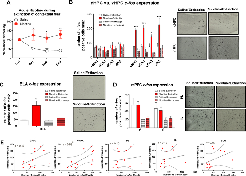
Figure 1. Acute nicotine administration during contextual fear extinction increases vHPC and BLA c-fos expression.

A. Normalized %freezing scores across initial testing and 3 extinctions sessions (n=6 per group). B. Acute nicotine increased the number of c-fos IR cells within the vHPC and vCA1, VCA3, and vDG but did not affect c-fos expression in the dHPC and its subregions. Right panel shows representative c-fos immunohistochemistry images from dHPC and vHPC of the Saline-Extinction and Nicotine-Extinction groups. C. BLA c-fos expression was increased in the group that received acute nicotine during contextual fear extinction. Right panel shows representative c-fos immunohistochemistry images from BLA of the Saline-Extinction and Nicotine-Extinction groups. D. Acute nicotine did not affect IL or PL c-fos expression, but extinction training increased c-fos expression in these brain regions. Right panel shows representative c-fos immunohistochemistry images from IL and PL of the Saline-Extinction and Nicotine-Extinction groups. E. Correlation plots between dHPC, vHPC, PL, IL, and BLA c-fos IR cell numbers and normalized %freezing scores for each subject. Error bars show standard error of the mean. *p<0.05; **p<0.01; ***p<0.001 compared to Saline-Extinction controls.
