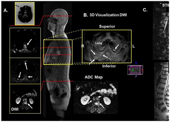
Figure 2.
Demonstration of WB-MRI on a 66-year-old man with metastatic prostate cancer. A. Axial diffusion imaging through the thorax and pelvis shows multiple metastatic lesions in the ribs, sternum, and vertebrae. B. 3D visualization of the thorax showing the metastatic lesions in the chest. C. T2 and STIR imaging of the spine to correlate the metastatic lesions with the WB-MRI.

