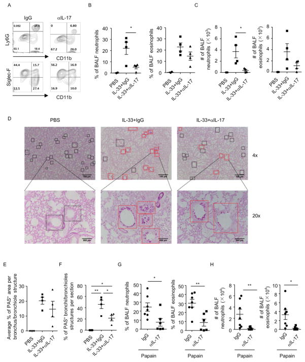
Figure 5. IL-33-driven IL-17 production plays a pathogenic role in lung inflammation.
Lung inflammation was induced in Rag1−/− mice by IL-33 or papain. Mice were injected with anti-IL-17 antibody or control IgG during induction of lung inflammation. BALF cells were counted and analyzed by flow cytometry. Data are representative of two independent experiments. (A–C and G–H) Expression of CD11b, Ly6G and Siglec-F in BALF cells gated on CD45+ cells was analyzed by flow cytometry. (B and G) Percentages of BALF neutrophils (CD11b+Ly6G+) and eosinophils (CD11b+SiglecF+) gated on CD45+ cells from indicated groups were shown. (C and H) Cell numbers of BALF neutrophils and eosinophils were shown. (D) Paraffin-embedded lung sections from indicated groups were stained with periodic acid-schiff (PAS). Magnifications are 4× and 20× respectively. Black squares indicated bronchi/bronchioles structures. Red squares indicated PAS+ bronchi/bronchioles structures. (E) Average percentages of PAS+ area in individually analyzed bronchioles/bronchi from indicated groups were shown. (F) Percentages of PAS+ bronchioles/bronchi among all bronchioles/bronchi per section from indicated groups were shown. (B, C, E–H) Data are means±SEM.

