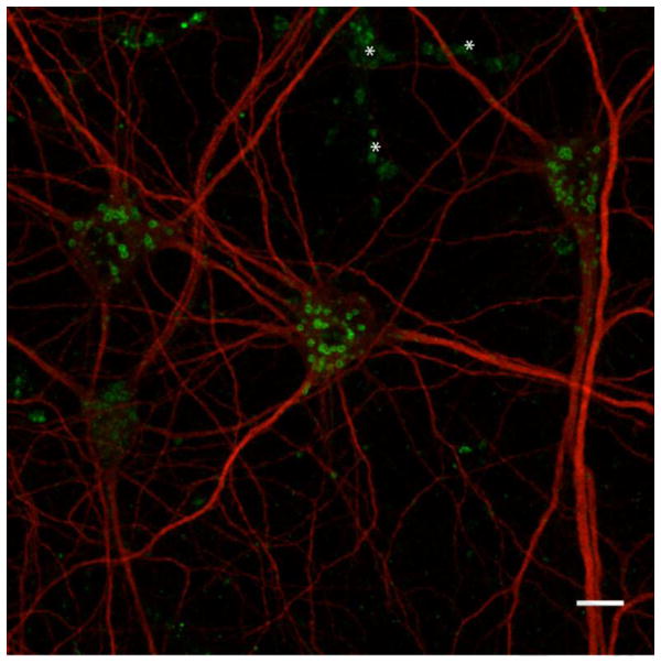
Figure 2.
Confocal image of lysosomes (LAMP1 signal, green) in mouse cortical neurons in culture along with MAP2B staining (red) which labels microtubules of neuronal dendrites. The asterisks indicate the position of non-neuronal lysosome signals arising from underlying glial cells. Although LAMP1 staining is commonly used to define lysosome distribution, due to the close relationship between lysosomes and other organelles of the secretory and endocytic pathways (Figure 1), the use of antibodies against multiple lysosomal proteins, including luminal hydrolases, is advisable for the definitive lysosome identification (Scale bar = 10 μm).

