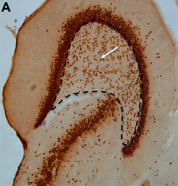
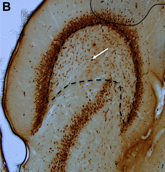
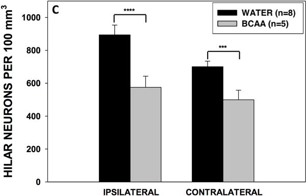
Figure 7. Hippocampal neuron counts.


(A) Section stained with NeuN demonstrating neurons within the dentate hilus (borders outlined with dashed lines) in a representative control and (B) branched-chain amino acid (BCAA) rat. The white arrows point to a stained neuron. (C) Rats in the branched-chain amino acid vs. control group had 36% fewer neurons in the dentate hilus of the hemisphere ipsilateral (p<0.0001) to the methionine sulfoximine infusion and 29% fewer neurons contralateral (p<0.001) to the infusion. Data is presented as mean number of cells per 100 mm3 ± SEM. ****p < 0.0001, ***p < 0.001
