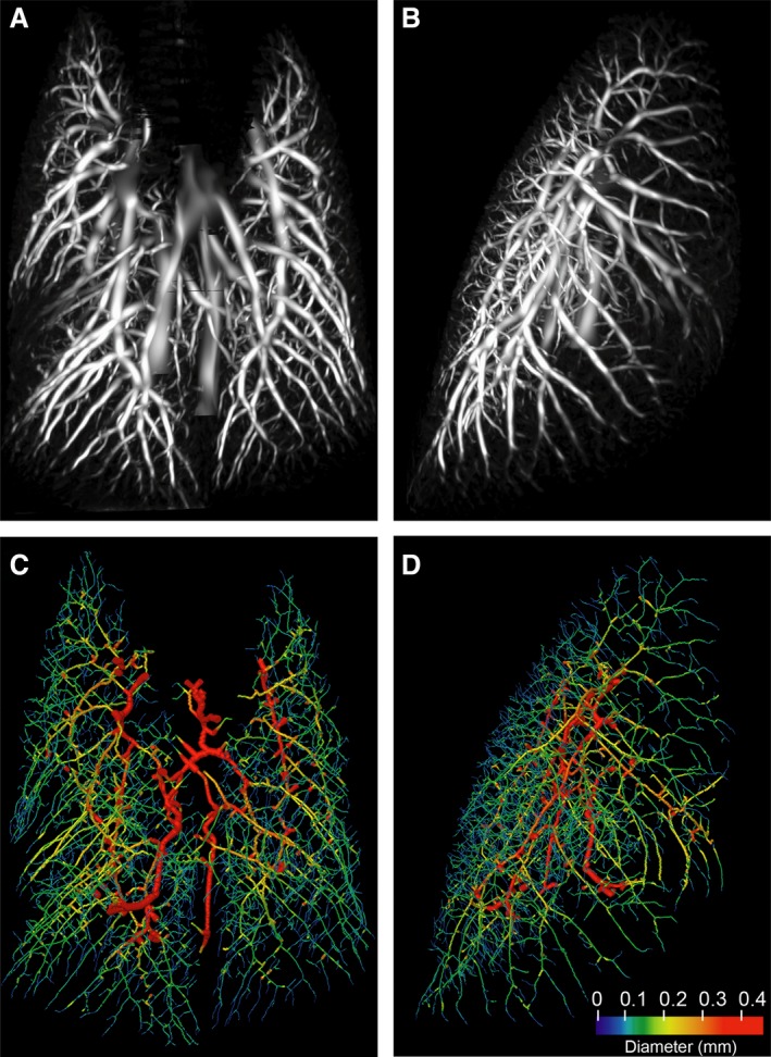
Figure 1.

Murine pulmonary vasculature derived from a CT scan without intravenous contrast. A healthy 8 week old BALB/c mouse was scanned once using dynamic x‐ray imaging and end‐inspiration images were segmented as described under Methods. (A, B) Probability field of the filtered image, as a maximum intensity projection (A, frontal; B, lateral); (C, D) Vessel diameters mapped to the centerline tree for the same animal/scan (C, frontal; D, lateral). Branch thickness is reduced by a factor of 6.8 for clarity.
