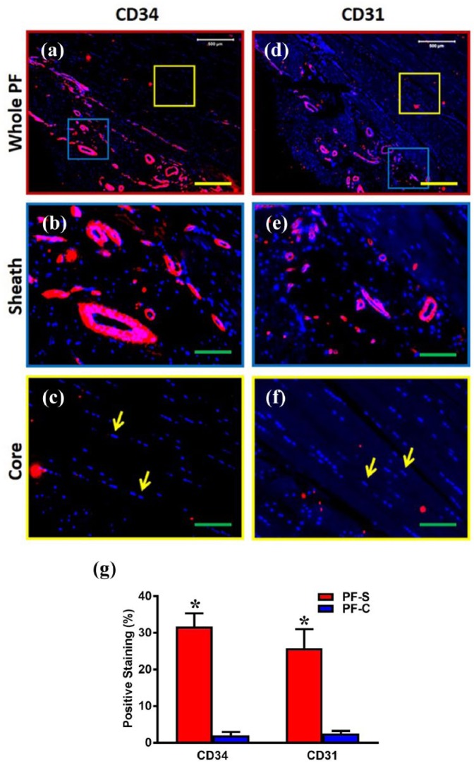
Figure 2.
The expression of endothelial cell markers, CD34 and CD31, is much higher in the sheath compared to the core in human PF tissue. The immunostaining on CD34 ((a)–(c)) and CD31 ((d)–(f)) shows that human PF tissue has sheath (blue box in (a), (d)) and core parts (yellow box in (a), (d)). Enlarged images of the blue box areas in (a) and (d) show that sheath tissue has a crosslink network of collagen fibers with many blood vessel–like tissues positively stained by CD34 and CD31 (red in (b), (e)). Enlarged images of the yellow box areas in (a) and (d) show elongated cells (yellow arrows in (c), (f)) stay in the core part with well-organized collagen fibers. There is no blood vessel–like tissue found in core part and very few cells are positively stained by CD34 and CD31 ((a), (c), (d), (f)). Red color represents areas positively stained with CD34 and CD31 and blue is stained nuclei. Semi-quantification shows significantly high staining for CD34 and CD31 in PF-S compared to PF-C cells (g). Yellow bars: 500 µm, green bars: 125 µm. *p < 0.01.

