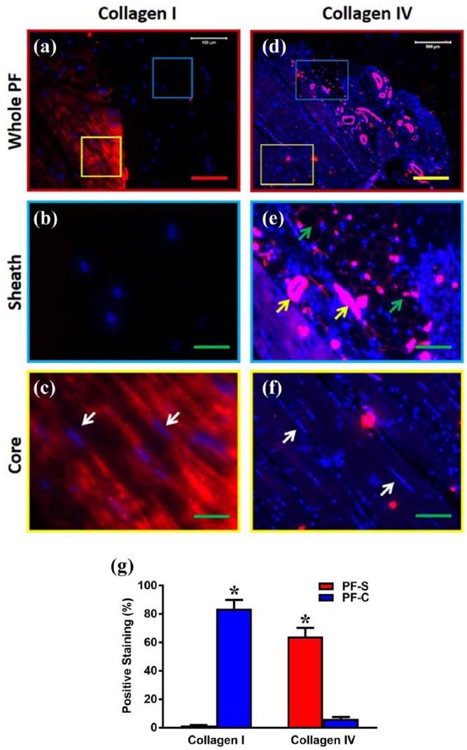
Figure 3.
The differential expression of collagen type I and collagen type IV in the sheath and core parts of human PF tissue. Immunostaining analysis shows that human PF has sheath (blue box in (a), (d)) and core parts (yellow box in (a), (d)). The enlarged image of the blue box area in (a) shows that sheath tissue has a crosslink network of collagen fibers negatively stained by collagen type I (b), while the enlarged image (e) of the blue box area in (d) shows that sheath tissue has a crosslink network of collagen fibers (green arrows in (e)) with many blood vessel–like tissues (yellow arrows in (e)) positively stained by collagen type IV. The enlarged image of the yellow box area in (a) shows elongated cells (white arrows in (c)) stay in the core part with well-organized collagen fibers positively stained with collagen I (c), while an enlarged image (f) of the yellow box area in (d) shows elongated cells (white arrows in (f)) stay in the core part with well-organized collagen fibers negatively stained with collagen IV. Semi-quantification shows significantly high collagen I in PF-C compared to PF-S and significantly high collagen IV in PF-S compared to PF-C cells (g). Red bar: 100 µm, yellow bar: 500 µm; green bars: 125 µm. *p < 0.01.

