Fig. 5.
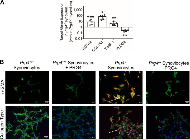
Gene expression of α-smooth muscle actin (ACTA2), collagen type I (COL1A1), tissue inhibitor of metalloproteinase-1 (TIMP-1), and procollagen-lysine, 2-oxoglutarate 5-dioxygenase 2 (PLOD2) in synovial tissues isolated from Prg4+/+ and Prg4−/− mice and immunocytostaining of α-smooth muscle actin (α-SMA) and collagen type I in Prg4+/+ and Prg4−/− synoviocytes and impact of human synoviocyte PRG4 treatment. A: ACTA2, COL1A1, and TIMP-1 expression in Prg4−/− synovial tissues was higher than in Prg4+/+ synovial tissues. PLOD2 expression in Prg4−/− synovial tissues was lower than in Prg4+/+ synovial tissues. Each group contained 5 samples, with each sample generated by pooling synovial tissues from 3 mice. B: merged images depicting α-SMA and collagen type I protein immunostaining in isolated Prg4+/+ synoviocytes and Prg4−/− synoviocytes (bright orange) and counterstained with F-actin (green) and DAPI (blue). α-SMA and collagen type I staining was detected in Prg4−/− synoviocytes (white arrows), and no staining was detected in Prg4+/+ synoviocytes. α-SMA and collagen type I staining intensities were reduced by human synoviocyte PRG4 treatment for 24 h. *P < 0.001, **P < 0.01, ***P < 0.05.

