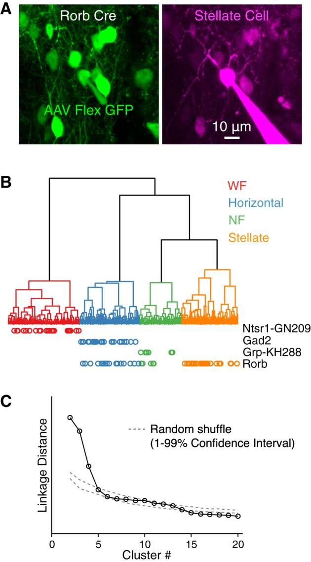
Fig. 1.

Nearly all stellate cells but not wide-field (WF) cells express Cre-dependent reporter in Rorb Cre mice. A: example green fluorescent protein (GFP)-expressing stellate cell recorded in superficial layers of the superior colliculus (sSC) in vitro after injection of AAV-Flex-ChR2-GFP in a Rorb Cre mouse. Note that some bright GFP-expressing cells are visible in the magenta channel, but the Alexa Flour 594 filling the recorded cell is not visible in the green channel. B: dendrogram representing, via vertical line lengths, the separation (linkage distance) of cells based on electrophysiological properties recorded in vitro. Vertical line ends at the bottom on the dendrogram represent individual cells (n = 878). Horizontal lines join similar cells into clusters. The 4 clusters shown in different colors correspond to the 4 cell types described by Gale and Murphy 2014. A subset of cells (n = 317) were recorded in 1 of 4 Cre lines (Table 1); cells from these experiments that expressed Cre-dependent reporter are indicated by colored circles below each cell’s position in the dendrogram. C: linkage distance between the 20 largest clusters in the dendrogram shown in A. Linkage distance is the increase in total within cluster variance (sum of squares of the Euclidean distance between each cell within a cluster and the cluster centroid in electrophysiological parameter space) that results from merging cluster N with cluster N-1. Dashed gray lines show the 1–99% confidence interval of linkage distances for clusters formed from randomly shuffled data. To generate randomly shuffled cluster data (1,000 repetitions), for each electrophysiological parameter, each cell was assigned a value for that parameter that was randomly drawn from the values of that parameter across all cells.
