Fig. 2.
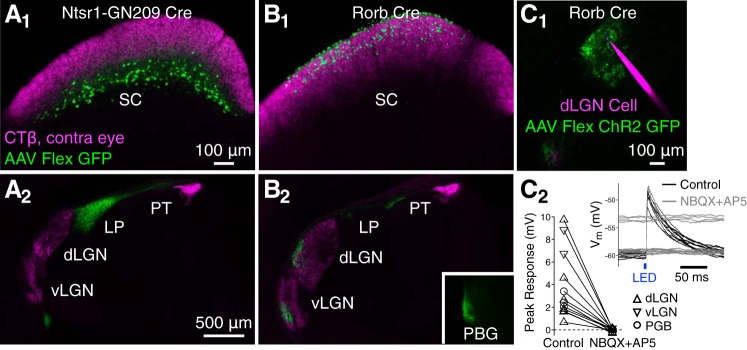
Stellate cells project to lateral geniculate nucleus (LGN) and parabigeminal nucleus (PBG) but not lateral posterior (LP) and are glutamatergic. All images are coronal sections with dorsal up and lateral left. A: green fluorescent protein (GFP)-expressing cells (green) at the injection site in the superior colliculus (SC; A1) and axons in thalamus (A2) after injection of AAV-Flex-GFP in a Ntsr1-GN209 Cre mouse. Retinal ganglion cell axons (magenta) were labeled via injection of cholera toxin subunit-β (CTβ) conjugated to Alexa 594 in the contralateral eye. GFP-expressing axons below ventral LGN (vLGN) in A2 are following the optic tract in route to the contralateral LP. PT, dorsal pretectum. B: same as in A for injections in a Rorb Cre mouse. C: synaptic responses to blue light (black and gray traces, C2) in a dorsal LGN (dLGN) cell (magenta, C1) recorded in vitro following AAV-Flex-ChR2-GFP (green, C1) injection in the sSC of a Rorb Cre mouse. Example traces were recorded before (black) and after (gray) bath application of the glutamate-receptor antagonists NBQX and AP5. Current injection was used to depolarize the cell for some trials after NBQX/AP5 application. The peak response to ChR2 stimulation, before and after NBQX/AP5 application, is shown in C2 for 7 dLGN (triangles), 2 vLGN (inverted triangles), and 3 PBG neurons (circles).

