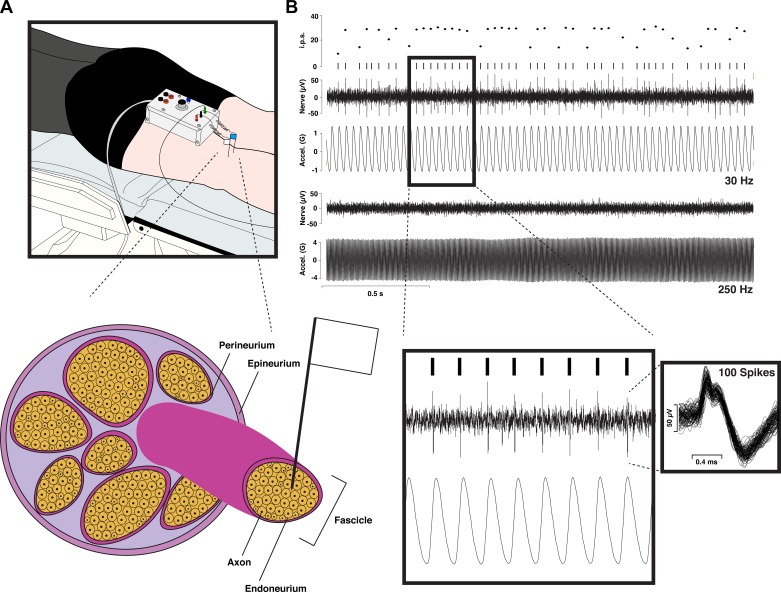
Fig. 1.
An illustration of the human microneurography technique. A, top: schematic of experimental setup for recording from the tibial nerve at the level of the knee (popliteal fossa). Two tungsten microelectrodes are inserted percutaneously, with one serving as the reference electrode inserted beneath the skin near the nerve and the other serving as the active electrode, which gets inserted into the nerve. Bottom, schematic of a peripheral nerve, showing the active electrode’s placement into an individual nerve fascicle, right up next to a single axon (i.e., intrafascicular extracellular recording). B: sample recording from a fast-adapting type I (FAI) afferent showing, from top to bottom, the instantaneous firing rate (i.p.s., impulses per second), raster plot, raw neurogram (Nerve), and vibrator acceleration (Accel.) for the case of 30- and 250-Hz vibration. As expected based on the FAI bandwidth, this unit codes precisely for the 30-Hz vibration with a phase-locked 30-Hz spike train but fails to be activated by the 250-Hz stimulation. Inset left: sample of phase-locking in the FAI response with the timescale expanded. Inset right: 100 overlaid spikes (note: the double-peaked action potential morphology indicates that the microelectrode has not caused conduction blockage; see Inglis et al. 1996).

