
Fig. 7.
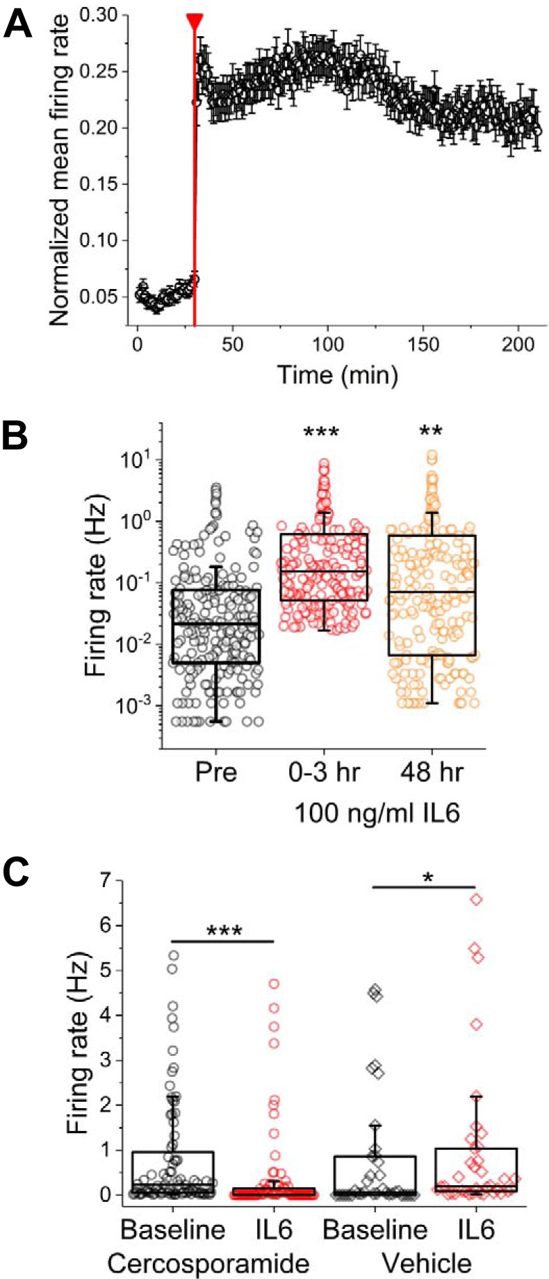
Spontaneous sensory neuron activity is significantly increased with short- and long-term incubation with IL-6. A: normalized mean firing rate for electrode subset found to be responsive to IL-6 (41% of previously active electrodes). Red line indicates addition of 100 ng/ml IL-6. B: firing rates before (Pre), immediately following (0–3 h), and 48 h following IL-6 addition. Symbols represent data from single electrodes. C: firing rates before and immediately following IL-6 addition in the presence of vehicle (water) or MAPK-interacting kinase 1/2 inhibitor cercosporamide. *P < 0.05, **P < 0.01, and ***P < 0.001.