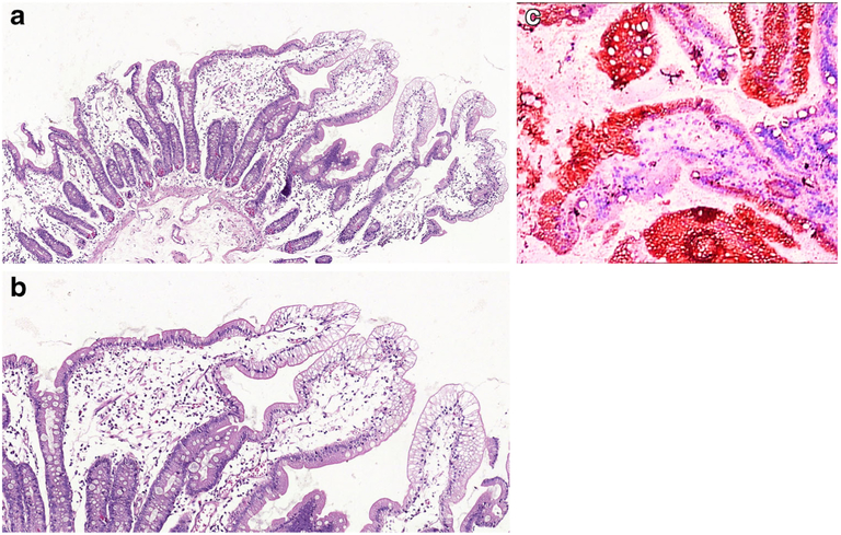
Fig.1.
a Duodenal mucosa from a 4-year-old boy with ABL. There is surface epithelial vacuolation in otherwise normallooking duodenal mucosa with preserved villus/crypt architecture (H&E; ×40). b Vacuolated appearance of surface enterocytes (H&E; ×200). c Lipid droplets in the enterocyte cytoplasm (Oil Red O; ×200)

