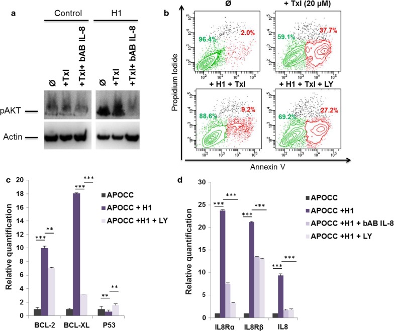
Fig. 4.
a Phospho-AKT western blot. APOCC cells pre-incubated or not with H1 with or without bAB IL-8 for 24 h were treated with Taxol (20 µM). Phospho-AKT was assessed by western blot. b Apoptosis assay. APOCC cells pre-incubated or not with H1 with or without an Akt inhibitor (LY294002) for 24 h were treated with Taxol (20 µM). Apoptosis was evaluated by flow cytometry using an apoptosis array. Live cells are represented in green and apoptotic cells in red. c The relative quantification of apoptosis genes was performed by real-time qPCR on APOCC before or after treatment with H1 or H1 + LY294002. Relative transcript levels are represented as the log10 of ratios between the two subpopulations of their 2−ΔΔCp real-time PCR values. d The relative quantification of IL-8 and IL-8 receptor genes was performed by real-time qPCR on APOCC before or after treatment with H1, H1 bAB IL-8 or H1 + LY294002. Relative transcript levels are represented as the log10 of ratios between the two subpopulations of their 2−ΔΔCp real-time PCR values

