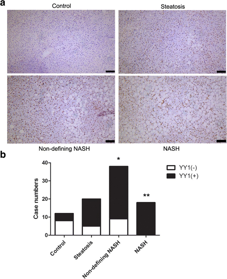
Fig. 3.

Immunohistochemistry detection of YY1 for NAFLD at different stages (a) Immunohistochemistry analysis was performed to detect the expression of YY1 in the control, steatosis, non-defining NASH, and NASH groups.Scale bar, 100 μm. b The cases positive or negative for YY1 expression were analyzed. *p < 0.05 compared with control group, **p < 0.01 compared with control group
